41 how to label a gel electrophoresis image
CHAPTER 10 Flashcards | Quizlet Please label the images to review the process of polymerase chain reaction and how its products can be analyzed using gel electrophoresis. Match the components of a typical PCR reaction with the function they serve. ... Please label the images to review the process of screening bacterial clones for those containing a donor gene. Other sets by ... ImageJ for Editing & Labelling PCR Gel Image - YouTube This Tutorial is all about how to quickly Edit & Label PCR Gel Image Using ImageJ software. Presented by - Elvis SamuelJoin Our Telegram Channel for free Sof...
How to make a gel image using Powerpoint - YouTube A quick tutorial on how to make a reasonably polished figure using an image of a gel using Powerpoint. There are certainly more professional ways of doing th...

How to label a gel electrophoresis image
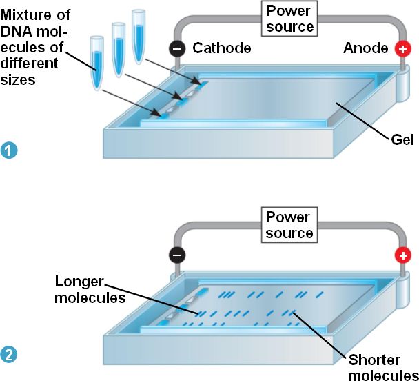
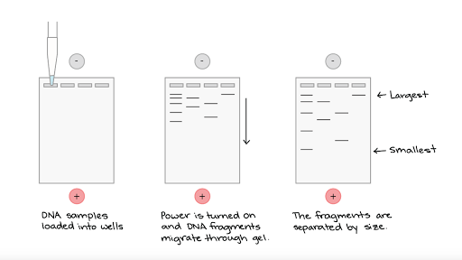
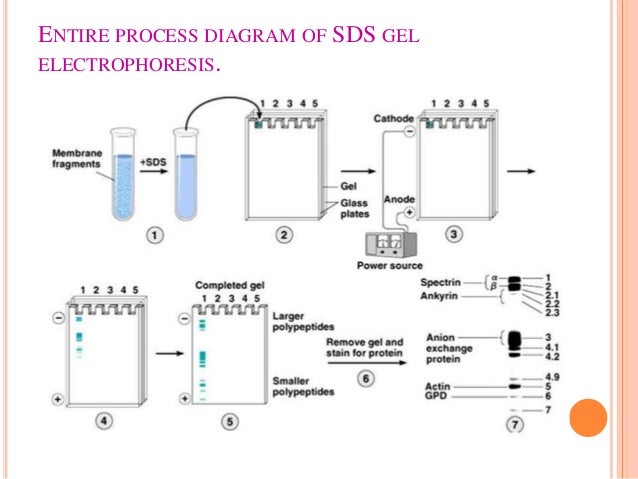
How to Interpret DNA Gel Electrophoresis Results - GoldBio During gel electrophoresis, you may have to load uncut plasmid DNA, digested DNA fragment, PCR product, and probably genomic DNA that you use as a PCR template into the wells. Your digested DNA fragment is a digested PCR product. The next step is to identify those bands to figure out which one to cut. Gel Electrophoresis. Lane 1: DNA Ladder. New Microsoft Word Document (4).docx - Gel electrophoresis... - Course Hero Gel electrophoresis A technique used to separate DNA fragments and other macromolecules by size and charge. Key points: Gel electrophoresis is a technique used to separate DNA fragments according to their size. DNA samples are loaded into wells (indentations) at one end of a gel, and an electric current is applied to pull them through the gel. DNA fragments are negatively charged, so they move ... Detection and quantitation of radiolabeled proteins and DNA in ... - PubMed This appendix presents procedures for visualizing and quantitating radiolabeled proteins or DNA separated by polyacrylamide gel electrophoresis or affixed to filter membranes. Autoradiography is the most common method by which this is accomplished; X-ray film is the traditional recording medium. Fil …
How to label a gel electrophoresis image. Figure legends Figure 1: Agarose gel electrophoresis (2% agarose) of ... Figure legends Figure 1: Agarose gel electrophoresis (2% agarose) of PCR amplified products using species-specific PCR primer sets. Lanes 1-17 are examined Salmonella isolates. Lanes 1-6 from... How to quantify each band in gel electrophoresis? - ResearchGate you can do an analytical curve in a 1d gel, with known amounts of bsa for example, use photoshop to quantify the pixels (the curve would be pixels x protein mass you applied for each well) and then... Analyzing gels and western blots with ImageJ - lukemiller.org This version of the tutorial was created using ImageJ 1.42q on a Windows 7 64-bit install. 1. Open the image file using File>Open in ImageJ. 2. The gel analysis routine requires the image to be a gray-scale image. The simplest method to convert to grayscale is to go to Image>Type>8-bit. Your image should look like Figure 1. Figure 1. DNA Ladders (1 kb, 1 kb plus, 100 bp, 100 bp plus) and Uses 1 kb ladder can be bought commercially at various concentrations, but the recommended load for an electrophoresis run is 0.5 µg (5µl). These can be used in either agarose or in polyacrylamide gels with the concentration of gel at 0.75% to 1%. These ladders come with different tracking dyes like bromophenol blue, xylene cyanol FF.
Detection and quantitation of radiolabeled proteins and DNA in ... - PubMed This appendix presents procedures for visualizing and quantitating radiolabeled proteins or DNA separated by polyacrylamide gel electrophoresis or affixed to filter membranes. Autoradiography is the most common method by which this is accomplished; X-ray film is the traditional recording medium. Fil … New Microsoft Word Document (4).docx - Gel electrophoresis... - Course Hero Gel electrophoresis A technique used to separate DNA fragments and other macromolecules by size and charge. Key points: Gel electrophoresis is a technique used to separate DNA fragments according to their size. DNA samples are loaded into wells (indentations) at one end of a gel, and an electric current is applied to pull them through the gel. DNA fragments are negatively charged, so they move ... How to Interpret DNA Gel Electrophoresis Results - GoldBio During gel electrophoresis, you may have to load uncut plasmid DNA, digested DNA fragment, PCR product, and probably genomic DNA that you use as a PCR template into the wells. Your digested DNA fragment is a digested PCR product. The next step is to identify those bands to figure out which one to cut. Gel Electrophoresis. Lane 1: DNA Ladder.
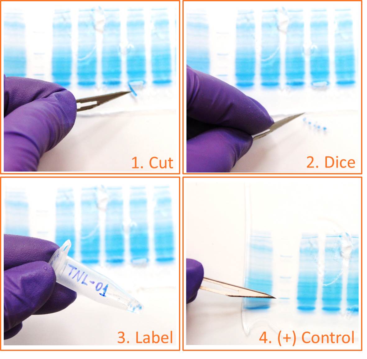
in-gel digestion | Proteomics and Mass Spectrometry Core Facility

Protein Mass Spectrometry Made Simple | American Society of Nephrology
How can tools of molecular biology be used to compare DNA of two ...
Gel electrophoresis (article) | Khan Academy
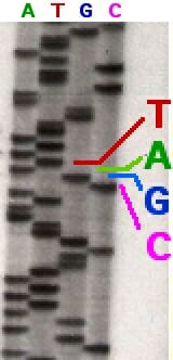
DNA Sequencing - Definition, Methods & Examples | Biology Dictionary
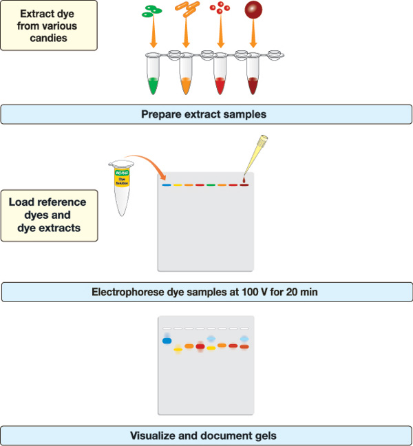
IDEA Kit — Inquiry Dye Electrophoresis Activity | Life Science ...

DNA fragment size and quantities in gel | Download Table

Mini Dual Vertical Electrophoresis Unit – TARSONS
Gel electroporosis
Post a Comment for "41 how to label a gel electrophoresis image"