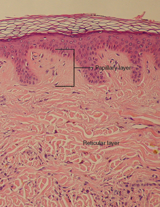
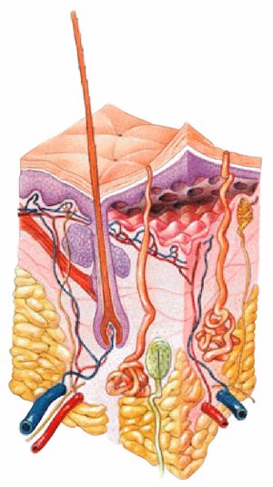
45 label the skin structures and areas indicated in the accompanying diagram of skin
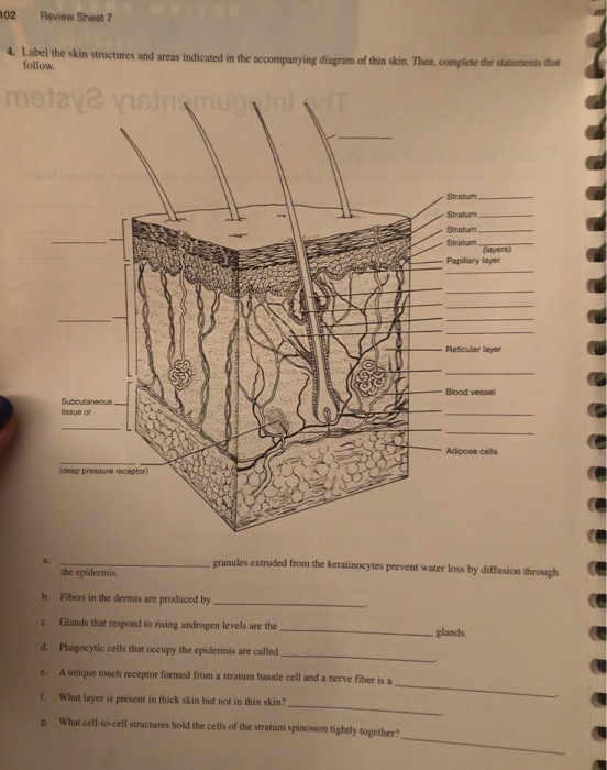
Solved > 13.Write the name of each tissue type in:1608366 ... - ScholarOn 4.Label the skin structures and areas indicated in the accompanying diagram of skin.... 5.What substance is manufactured in the skin (but is not a secretion) to play a role elsewhere in the body? ___________ 6.Some injections hurt more than others.... Solved 4. Label the skin structures and areas indicated in - Chegg Stratum Popilary layer Reticular layer Blood vessel tissue or Adpose cels (deep pressure receptor granules contain glycolipids that prevent water loss from the skin. b. Fibers in the dermis are produced by c. Question: 4. Label the skin structures and areas indicated in the accompanying diagram of thin skin.
Neufert 4th Edition | PDF | Elevator | Roof - Scribd ~ p pressed connection 50 , l 40 change of nominal diameter, pipe ventilator, thermometer e.g. from 0 50 to 0 40 l>l

Label the skin structures and areas indicated in the accompanying diagram of skin
Brain lab - lab - Answer the following questions and locate the ... Complete the diagram on Broca's areas. a. Temporal- b. Occipital- c. Frontal-d. Parietal-(6) Locate the brainstem on the lab specimen. Examine the model of the brainstem and locate each component and state the function of each. Label these structures on the accompanying diagram. a. Medulla oblongata-b. Pons- c. Midbrain- Solved tive tissue 4. Label the skin structures and areas - Chegg Label the skin structures and areas indicated in the accompanying diagram of thin skin. Then, complete the statements that follow. Weisshaft Stratum opidamist Stratum Stratum Stratum Papilary layer Dermis Reticular layer ascectors allmusde Encrine Sweet blond Dermal Vascular plexus pensery neare fiber Blood vessel Subcutaneous tissue or 4 Label the skin structures and areas indicated in the accompanying ... 4. Label the skin structures and areas indicated in the accompanying diagram of thin skin. Then, complete the statements that follow.
Label the skin structures and areas indicated in the accompanying diagram of skin. PDF Integumentary Review Sheet - Home - Holly H. Nash-Rule, PhD Name Lab Time/Date The Integumentary System Basic Structure of the Skin 1. Complete the following statements by writing the appropriate word or phrase on the correspondingly numbered blank: The two basic tissues of which the skin is composed are dense irregular 1. connective tissue, which makes up the dermis, and 1 , which forms the epidermis. Skin Diagram with Detailed Illustrations and Clear Labels - BYJUS Skin Diagram The largest organ in the human body is the skin, covering a total area of about 1.8 square meters. The skin is tasked with protecting our body from external elements as well as microbes. Interesting Note: PDF Label The Skin Structures And Areas Indicated Label The Skin Structures And Areas Indicated Therapeutic Injections for Pain Management Types of April 28th, 2018 - This article focuses on the use of therapeutic injections see the image below to treat acute and chronic pain syndromes Discussion of this topic begins with an overview of regional anesthesia Houston Community College Label the skin structures and areas indicated in the accompanying diagram of thin skin. Then, complete the statements that follow. Subcutaneous tissue or (deep pressure receptor) the epidermis. Stratum Stratum Stratum Stratum (layers) Papillary layer Reticular layer Blood vessel Adipose cells
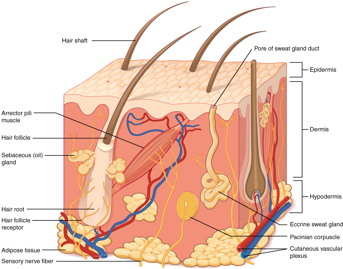
Solved: Chapter E7 Problem 4E Solution | Masteringa&p - Chegg Step 1 of 3 Keratinocytes also called keratin cells are the cells of epidermis. Lamellated granules extruded from these keratinocytes prevent the loss of water by diffusion through the epidermis. Thus maintain the sufficient water content in the body. 4. Label the skin structu - YUMPU 4. Label the skin structures and areas indicated in the accompanying diagram of skin. epidermis. dermis. Subcutaneous. tissue or. hypodermis. Pacinian corpuscle (deep pressure receptor) 5. What substance is manufactured in the skin (but is not a secretion) to play a role elsewhere in the body? The skin is the site of vitamin D synthesis for the ... PDF The Integumentary System - Holly H. Nash-Rule, PhD Label the skin structures and areas indicated in the accompanying diagram of thin skin. Then, complete the statements that follow. a. Lamellar granules contain glycolipids that prevent water loss from the skin. b. Fibers in the dermis are produced by fibroblasts . c. Label Skin Diagram Printout - EnchantedLearning.com epidermis - the outer layer of the skin. hair follicle - a tube-shaped sheath that surrounds the part of the hair that is under the skin. It is located in the epidermis and the dermis. The hair is nourished by the follicle at its base (this is also where the hair grows). hair shaft - The part of the hair that is above the skin.
. 4. Label the skin structures and areas indicated in the... Sweat gland are pound throughout the skin but are more numerous in area such as the soly op the Reet, palm of the hand , armpits and groins, The ectine sweat glad controled by the sympathetic Nervous system which regulate the body temperature e Shot on OnePlus By Herowl Label the Skin | Biology Diagram | Quizlet Epidermis. layer of the skin that is continually being shed to make room for new skin. Dermis. layer of the skin that contains nerves, vessels, hair follicles, glands, and muscles. Hypodermis. layer of the skin that is used for fat storage. Sweat Pore. opening in the skin that sweat comes out of. Hair Follicle. (PDF) LIBRO PARA COLOREAR NETTER - Academia.edu Background: The aim of our study was to examine the effect of mild maternal hypothyroidism on the apoptosis of the oocytes in the ovaries of rats in the early postnatal period during formation of oocytes and follicles. Solved 104 Review Sheet 7 4. Lobal the bi Label the skin - Chegg Lobal the bi Label the skin structures and areas indicated in the accompanying diagram of thin skin. Then, complete the statements that follow -hair shaft Stratum Stratum Stratum Stratum epidermis Papillary layer Permis Reticular layer Blood vessel Subcutaneous tissue or Hypodermish Adipose cells (deep pressure receptor) granules
CFTR and Cystic Fibrosis: A Need for Personalized Medicine ...
Anatomy and Physiology Homework Chapter 6 Flashcards | Quizlet Study with Quizlet and memorize flashcards containing terms like Label the parts of the skin and subcutaneous tissue. -Blood Capillaries -Piloerector muscle -Dermal papilla -Hair bulb -Sensory nerve fibers -Tactile corpuscle -Hair follicle -Sebaceous gland, Label the parts of the skin and subcutaneous tissue. -Hypodermis -Sweat pores -Dermis -Hairs -Cutaneous blood vessels -Epidermis -Sweat ...
Quentin Bone. 17 August 1931â•fl6 July 2021
13. CHAPTER XIII. INTEGUMENTARY SYSTEM_LAB.docx - ROJO,... Hypodermis: it consists largely of fat. It provides the main structural support for the skin, as well as insulating the body from cold and aiding shock absorption. It is interlaced with blood vessels and nerves.
The Integumentary System
PDF Integumentary Review Packet Key - Home - Buckeye Valley Integumentary Review Packet Key - Home - Buckeye Valley
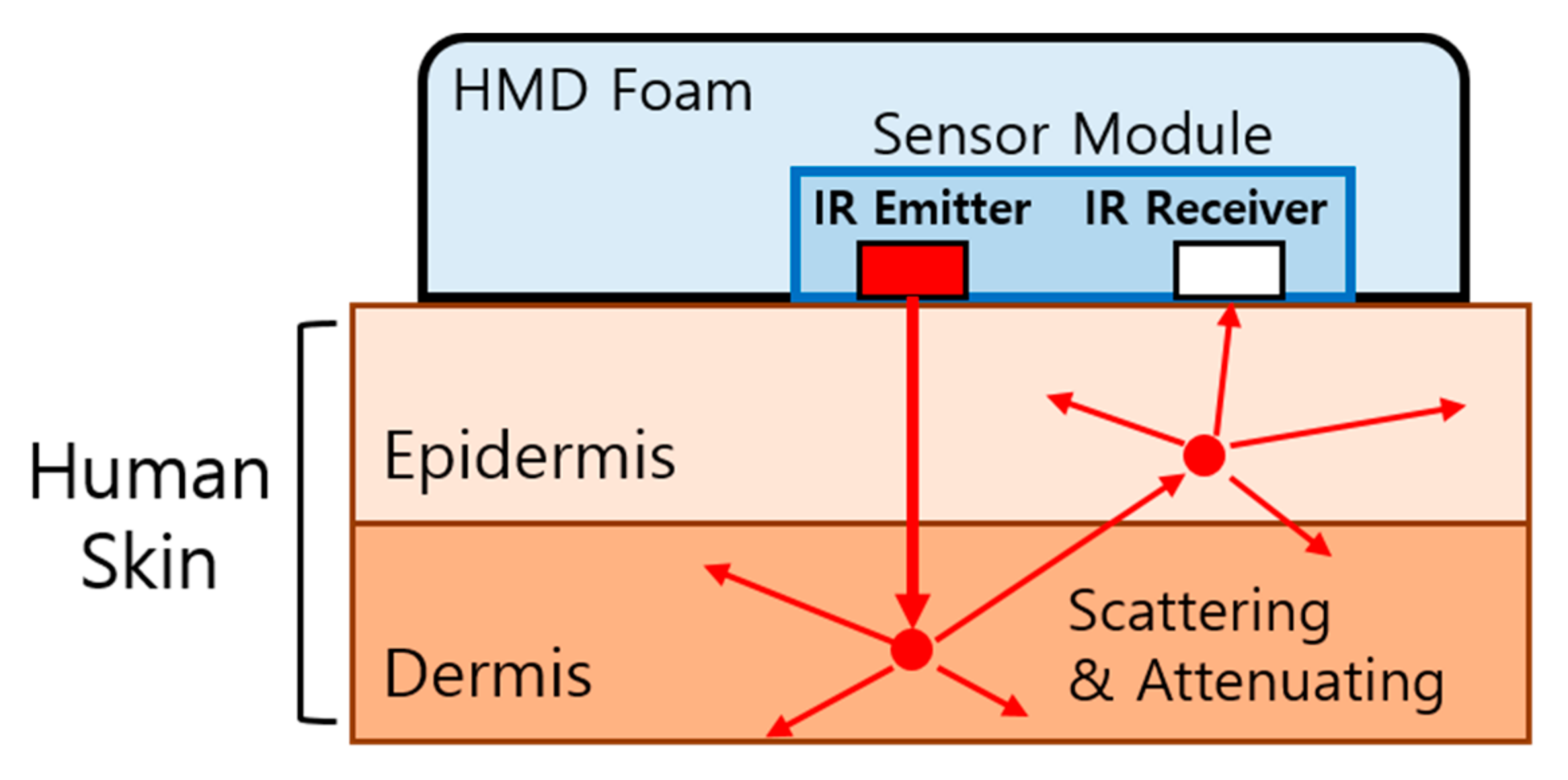
Sensors | Free Full-Text | Hands-Free User Interface for VR ...
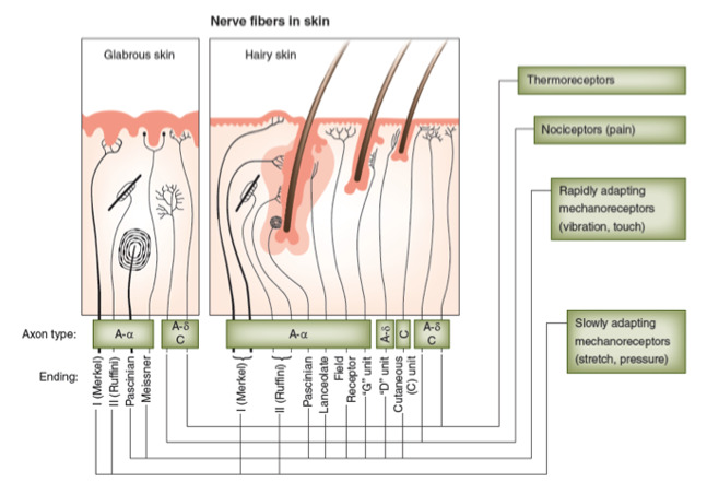
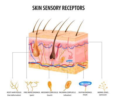
Solved > 14.Using the key, choose the best response to:1609438 ... 4.Label the skin structures and areas indicated in the accompanying diagram of thin skin. Then, complete the statements that follow. a. granules contain glycolipids tha... 5.What substance is manufactured in the skin and plays a role in calcium absorption elsewhere in the body? 6.List the sensory receptors found in the dermis...

A & P 1 Chapter 5 Flashcards | Quizlet
Solved C. Label the skin structure and areas indicated in - Chegg C. Label the skin structure and areas indicated in the accompanying diagram of skin D. Appendages of the Skin Using the key choices, respond to the following descriptions. (Some choices may be used more than once). Key: arrector pili muscle Cutaneous receptors Hair Sweat gland-apocrine Sweat gland-eccrine hair follicle nail sebaceous glands 1.
Diabetic Neuropathies - Endotext - NCBI Bookshelf
Botox ingredients list - gnsgz.autec-vlt.de Aug 02, 2017 · The star ingredient, XEP 30, a neuropeptide that's similar to Botox, eases creases and folds, leaving softened skin and smoother-looking lines. The area between the eyes (above the bridge of the nose). Injections here are indicated for the treatment of "11" lines, a common cosmetic issue (dosing varies). The corners of the eyes.

Solved 104 Review Sheet 7 4. Lobal the bi Label the skin ...
The Integumentary System_page2_answers.png - 4. Label the skin ... Label the skin structures and areas indicated in the accompanying diagram of thin skin. Then, complete The Integumentary System_page2_answers.png - 4. Label the... School South University, Savannah Course Title BIO 1012 Uploaded By Xtina0826 Pages 1 This preview shows page 1 out of 1 page. View full document End of preview.
5.1 Layers of the Skin – Anatomy & Physiology
Anatomy and Physiology 2e - 2e - Open Textbook Library Anatomy and Physiology 2e is developed to meet the scope and sequence for a two-semester human anatomy and physiology course for life science and allied health majors. The book is organized by body systems. The revision focuses on inclusive and equitable instruction and includes new student support. Illustrations have been extensively revised to be clearer and more inclusive. The web-based ...

The interaction of metals and the skin: The good, bad, and ...
Solved > 19.The three types of muscle tissue exhibit similarities ... 4.Label the skin structures and areas indicated in the accompanying diagram of thin skin. Then, complete the statements that follow. a. granules contain glycolipids tha... 5.What substance is manufactured in the skin and plays a role in calcium absorption elsewhere in the body? 6.List the sensory receptors found in the dermis...

Behind the Ice: The Archaeology of Nunatarsuaq, Southwest ...
Solved > 20.Label the tissue types illustrated here and on:1609441 ... 4.Label the skin structures and areas indicated in the accompanying diagram of thin skin. Then, complete the statements that follow. a. granules contain glycolipids tha... 5.What substance is manufactured in the skin and plays a role in calcium absorption elsewhere in the body? 6.List the sensory receptors found in the dermis...
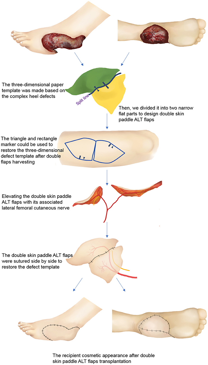
Frontiers | Reconstruction of Complex Soft Tissue Defects of ...
WebMD - Better information. Better health. The skin is the largest organ of the body, with a total area of about 20 square feet. The skin protects us from microbes and the elements, helps regulate body temperature, and permits the ...
5.1 Layers of the Skin – Anatomy & Physiology
03 Label the Cell Diagram | Quizlet Start studying 03 Label the Cell. Learn vocabulary, terms, and more with flashcards, games, and other study tools. ... cell diagram. 18 terms. lugo_janet. Sets found in the same folder. 03 Organelle Functions. 14 terms. muskopf1. 07 Cell Labeling. ... Hole's Human Anatomy and Physiology 13th Edition David N. Shier, Jackie L. Butler, Ricki Lewis ...

Biointerface Structural Effects on the Properties and ...
Optical Metasurfaces for Energy Conversion | Chemical Reviews Nanostructured surfaces with designed optical functionalities, such as metasurfaces, allow efficient harvesting of light at the nanoscale, enhancing light–matter interactions for a wide variety of material combinations. Exploiting light-driven matter excitations in these artificial materials opens up a new dimension in the conversion and management of energy at the nanoscale. In this review ...

Skin 1: the structure and functions of the skin | Nursing Times
Skin Structure (Labeling) Flashcards | Quizlet Where is Adipose Tissue Located on the skin structure? Sets with similar terms. Skin 1. 47 terms. lorianderson44 TEACHER. CHAPTER 10 (INTEGUMENT) 59 terms. emorrrry PLUS. Human Anatomy. 15 terms. hairstons. Integumentary Systems. 76 terms. Linnea_Eastburg11. Sets found in the same folder. Histology Lab Photo Quiz. 46 terms. robswatski TEACHER.
The Skin (Integumentary System)
Assignment 11 pg 104.pdf - 4. Label the skin structures and areas ... Label the skin structures and areas indicated in the accompanying diagramofthin skin. Then, complete the statements thatfollow. SubcutaneousJtissue or _l T~P-r
Skin Anatomy, Physiology, and Healing Process - Physiopedia
(PDF) Nanda NIC NOC | dwi adiyanto - Academia.edu Enter the email address you signed up with and we'll email you a reset link.
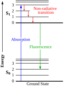
Fluorescence - Wikipedia
The Language of Anatomy - Holly H. Nash-Rule, PhD Indicate the following body areas on the accompanying diagram by placing the correct key letter at the end of each line. Key: a. abdominal b. antecubital c. brachial d. cervical e. crural f. femoral g. fibular h. bgluteal i. lumbar j. occipital k. oral l. popliteal m. pubic n. sural o. thoracic p. umbilical k o a p m f g e d j c i h l n Name _____
Solved NAME 7 The Integumentary System LAB TIME/DATE REVIEW ...
Solved > 4.Label the skin structures and areas indicated in:1609444 ... 4.Label the skin structures and areas indicated in the accompanying diagram of thin skin. Then, complete the statements that follow. a. granules contain glycolipids that prevent water loss from the skin. b. Fibers in the dermis are produced by . c. Glands that respond to rising androgen levels are the glands. d.

image086.jpg
4 Label the skin structures and areas indicated in the accompanying ... 4. Label the skin structures and areas indicated in the accompanying diagram of thin skin. Then, complete the statements that follow.
Peritoneal Dialysis Guidelines 2019 Part 1 (Position paper of ...
Solved tive tissue 4. Label the skin structures and areas - Chegg Label the skin structures and areas indicated in the accompanying diagram of thin skin. Then, complete the statements that follow. Weisshaft Stratum opidamist Stratum Stratum Stratum Papilary layer Dermis Reticular layer ascectors allmusde Encrine Sweet blond Dermal Vascular plexus pensery neare fiber Blood vessel Subcutaneous tissue or
Alternative Routes of Drug Administration (analgesia)
Brain lab - lab - Answer the following questions and locate the ... Complete the diagram on Broca's areas. a. Temporal- b. Occipital- c. Frontal-d. Parietal-(6) Locate the brainstem on the lab specimen. Examine the model of the brainstem and locate each component and state the function of each. Label these structures on the accompanying diagram. a. Medulla oblongata-b. Pons- c. Midbrain-

Assignment 11 pg 104.pdf - 4. Label the skin structures and ...
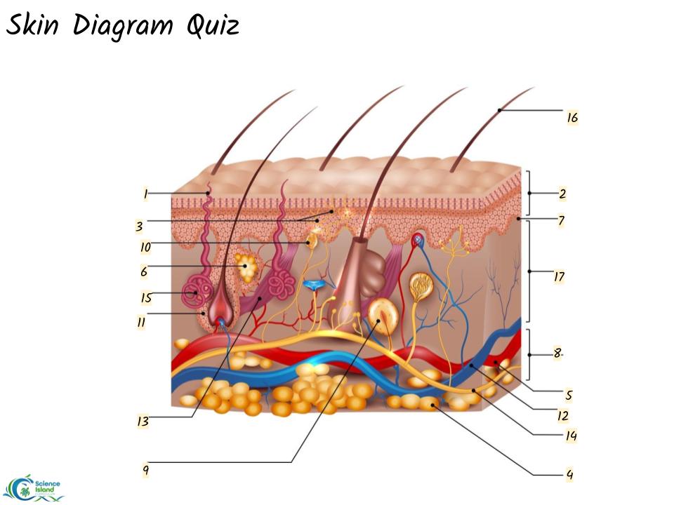
Skin Diagram Quiz | Science - Quizizz

Skin Diagrams & Quiz - Science Island
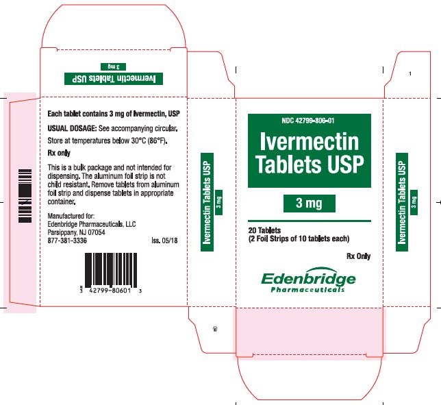
Ivermectin Tablets: Package Insert / Prescribing Information ...

ADDITIONAL ACTIVITY Direction: Label the skin structures and ...

Anatomy 2017- Unit 2 Label Parts of Skin Diagram Diagram ...

Assignment 11 pg 104.pdf - 4. Label the skin structures and ...

Human Physiology/Print Version - Wikibooks, open books for an ...
A New Concept of Static Rubber Gasket for Sealing Rough Surface

Fast, strong, and reversible adhesives with dynamic covalent ...

Label-Free SERS Analysis of Urine Using a 3D-Stacked AgNW ...
Carboplatin: Package Insert / Prescribing Information - Drugs.com
The Integumentary System

Recent Advances in Analytical Approaches for Glycan and ...
Normal Variation in the Anatomy, Biology, and Histology of ...

Polymeric Tissue Adhesives | Chemical Reviews
Review of deep learning: concepts, CNN architectures ...
Integumentary System - Physiopedia
Skin Anatomy - EnchantedLearning.com

Skin Structure (Labeling) Flashcards | Quizlet

Skin Cancer Treatment (PDQ®) - PDQ Cancer Information ...
TYPE HEADING HERE
BY ORDER OF THE SECRETARY OF THE AIR FORCE AIR FORCE PAMPHLET ...
5.1 Layers of the Skin – Anatomy & Physiology
Post a Comment for "45 label the skin structures and areas indicated in the accompanying diagram of skin"