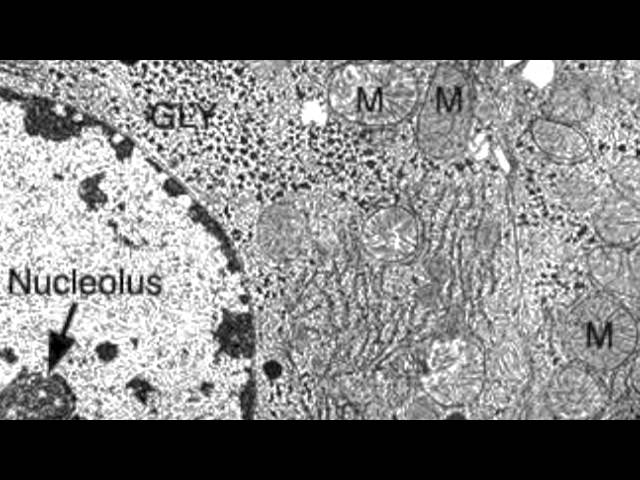
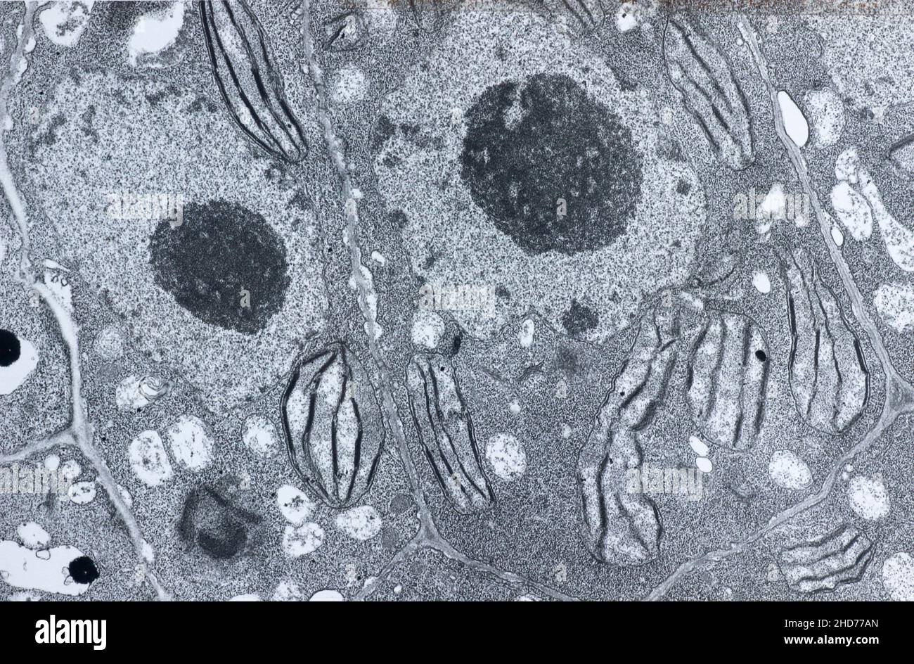
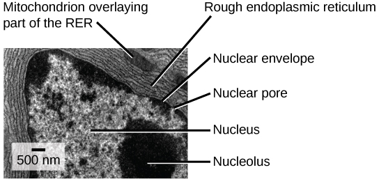
39 label the transmission electron micrograph of the cell
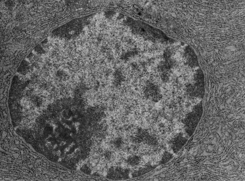
Transmission Electron Microscope (TEM) - Uses, Advantages and Disadvantages Advantages. A Transmission Electron Microscope is an impressive instrument with a number of advantages such as: TEMs offer the most powerful magnification, potentially over one million times or more. TEMs have a wide-range of applications and can be utilized in a variety of different scientific, educational and industrial fields. Solved Label the transmission electron micrograph based on - Chegg Expert Answer nucleus is the house of the genetic material which contains all the h … View the full answer Transcribed image text: Label the transmission electron micrograph based on the hints provided Mitochondrion Heterochromatin Plasma cell Nucleus Rough endoplasmic reticulum Nucleolus Previous question Next question
Label This Transmission Electron Micrograph / Microscopy Innovations ... Label the transmission electron micrograph of the nucleus. Transmission electron micrographs of hela cell sections labeled in . Label the transmission electron micrograph of the nucleus. Fluorescence microscopy in combination with tem and an ion beam analysis (iba, which allows the evaluation of the chemical elemental distribution) has allowed .

Label the transmission electron micrograph of the cell
Immune response: MedlinePlus Medical Encyclopedia Lymphocytes are a type of white blood cell. There are B and T type lymphocytes. B lymphocytes become cells that produce antibodies. Antibodies attach to a specific antigen and make it easier for the immune cells to destroy the antigen. T lymphocytes attack antigens directly and help control the immune response. Electron Micrographs of Cell Organelles | Zoology - Biology Discussion This is an electron micrograph of nucleus. (Fig. 17 & 18): (1) Nucleus was discovered by Brown (1831). (2) It is a characteristic entity of almost all eukaryotic cells except mammalian RBCs. (3) The nucleus is generally one but may also be two, four or many. Micro Quiz 2 (Chapters 3, 4) Flashcards | Quizlet transmission electron microscope confirming the 9 + 2 microtubule arrangement in a eukaryotic flagellum viewing ribosomes (20 nm) within a bacterial cell viewing a cross section of poliovirus (30 nm)viewing the layers of the gram-negative cell wall in cross section
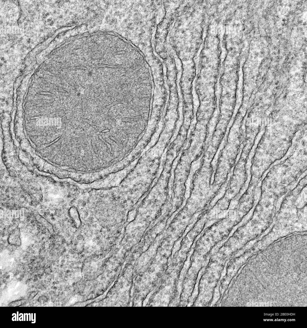
Label the transmission electron micrograph of the cell. Ultrasensitive detection of patulin based on a Ag+-driven one ... After the formation of stable Au-N bonds between AuNFs and g-C 3 N 4 nanosheets by chemical bonding, the typical transmission electron micrograph of the AuNFs/g-C 3 N 4 composite in Fig. 1 G shows that flower-like particles are fixed on a single nanosheets, indicating that AuNFs were successfully loaded onto the surface of g-C 3 N 4 nanosheets. Electron microscope - Wikipedia The original form of the electron microscope, the transmission electron microscope (TEM), uses a high voltage electron beam to illuminate the specimen and create an image. The electron beam is produced by an electron gun, commonly fitted with a tungsten filament cathode as the electron source. The electron beam is accelerated by an anode typically at +100 keV (40 to … Tour of The Cell and Microscopy Flashcards | Quizlet A transmission electron microscope profiles a thin section of a specimen. This EM shows a section through a tracheal cell, revealing its internal structure. In preparing the specimen, some cilia were cut along their lengths, creating longitudinal sections, while other cilia were cut straight across, creating cross sections. ABBREVIATION TEM Electron Micrographs - University of Oklahoma Health Sciences Center Figure 1 Micrograph of a nucleus. 1. Heterochromatin 2. Euchromatin 3. Nucleolus 4. Nucleolar associated chromatin 5. Nuclear envelope Figure 2 Micrograph of a portion of a nucleus: What is the round structure (approximately 3 1/2 inches in diameter) seen in the center of this micrograph? 1. Nucleolar associated chromatin 2.
Diagram Of Animal Cell As Seen Under Electron Microscope : Plant Cell ... A human lymphocyte white blood cell as seen under a transmission electron microscope cell theory the scientists schleiden and schwann observed plant and animal tissues under the microscope and. V, w, x and are the organelles in the animal cell while structure z can be found in the nucleus. ... plant cell seen under light microscope the cell as ... BacPROTACs mediate targeted protein degradation in bacteria - Cell 03-06-2022 · Small-molecule adaptors, BacPROTACs, redirect bacterial ClpCP protease to target neo-substrates in a highly specific manner and expand targeted protein degradation technology to … Junqueira's Basic Histology Text and Atlas, 14th Edition Junqueira's Basic Histology Text and Atlas, 14th Edition WHO plans to rename monkeypox over stigmatization concerns Aug 12, 2022 · Read More FILE – This image provided by the National Institute of Allergy and Infectious Diseases (NIAID) shows a colorized transmission electron micrograph of monkeypox particles (red) found ...
Fluorescence microscope - Wikipedia Fluorescence micrograph gallery A z-projection of an osteosarcoma cell, stained with phalloidin to visualise actin filaments. The image was taken on a confocal microscope, and the subsequent deconvolution was done using an experimentally derived point spread function. WHO plans to rename monkeypox over stigmatization concerns 12-08-2022 · LONDON (AP) — The World Health Organization says it’s holding an open forum to rename the disease monkeypox, after some critics raised concerns the name could be derogatory or have racist ... Solved Label the transmission electron micrograph of the - Chegg Expert Answer 100% (4 ratings) Explanation - Mitochondrion is filamentous or globular in shape, occur in variable numbers from a few hundred to few thousands in different cells. It … View the full answer Transcribed image text: Label the transmission electron micrograph of the mitochondrion. MicroRNA sequence codes for small extracellular vesicle ... Dec 22, 2021 · a, The experimental set-up and cell lines used in this study.b, PCA showing cellular (triangles) and sEV (circles) miRNA profiles for each cell type (n = 3–4).c, Venn diagram showing the number ...
Transmission electron micrographs of the ink-release vesicle ...
Electron microscope - Wikipedia An electron microscope is a microscope that uses a beam of accelerated electrons as a source of illumination. As the wavelength of an electron can be up to 100,000 times shorter than that of visible light photons, electron microscopes have a higher resolving power than light microscopes and can reveal the structure of smaller objects.

Transmission electron microscopy (TEM) imaging of nuclear ...
Looking at the Structure of Cells in the Microscope Determining the detailed structure of the membranes and organelles in cells requires the higher resolution attainable in a transmission electron microscope. Specific macromolecules can be localized with colloidal gold linked to antibodies. Three-dimensional views of the surfaces of cells and tissues are obtained by scanning electron microscopy.

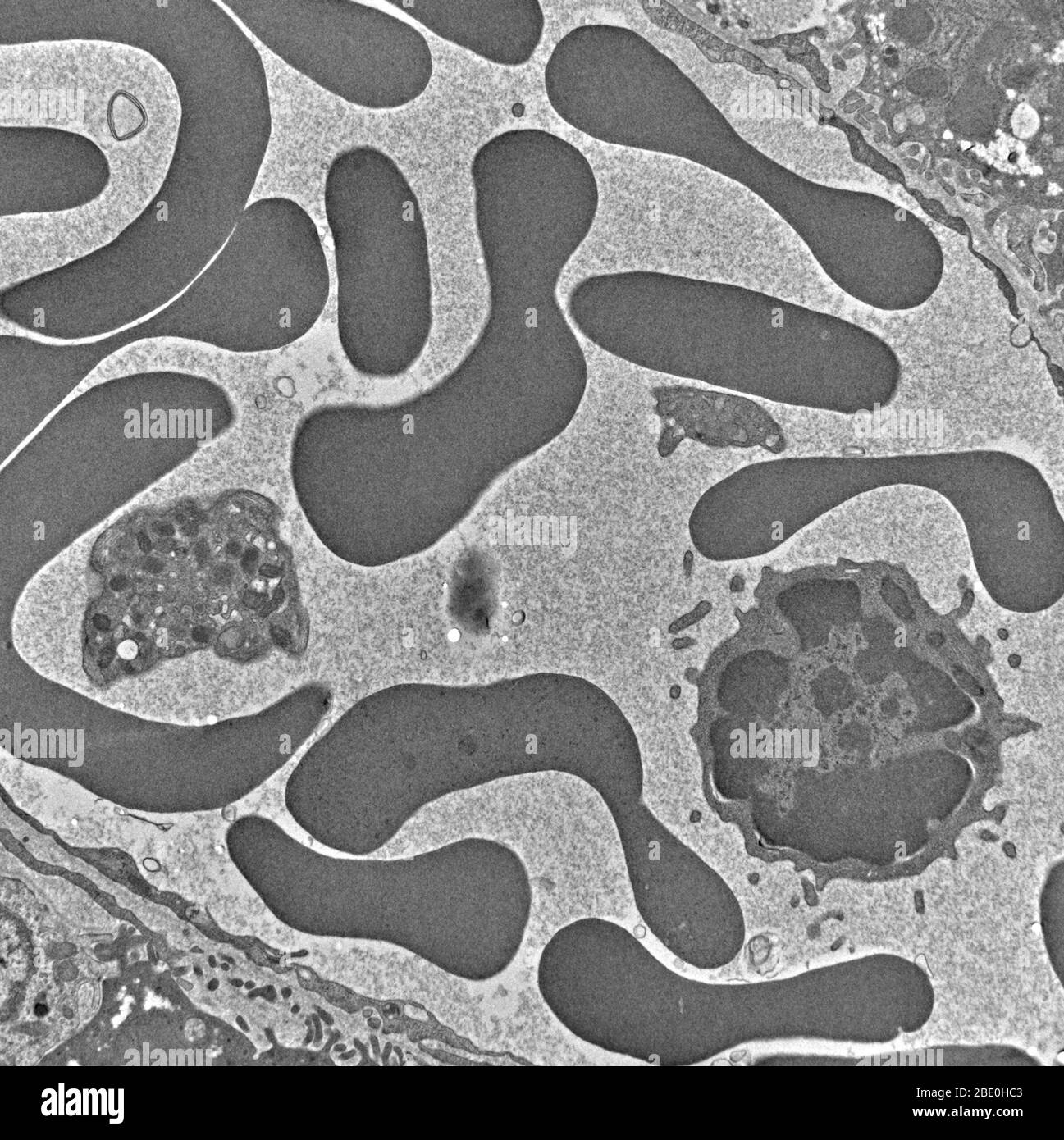
Transmission electron micrograph of mitochondrial and ...
Liquid-Cell Electron Microscopy of Adsorbed Polymers - PubMed Liquid-Cell Electron Microscopy of Adsorbed Polymers. Adv Mater. 2017 Nov;29 (41). doi: 10.1002/adma.201703555. Epub 2017 Sep 18.

Transmission electron microscopy (TEM) imaging of nuclear ...
Label This Transmission Electron Micrograph - Kaiden Brown Label the transmission electron micrograph of the nucleus. Label the transmission electron micrograph of the nucleus. Transmission electron microscopy (tem) is a microscopy technique in which a beam of electrons is transmitted through a specimen to form an image. Figures label this transmission electron micrograph ( 16, . CIN2003. Ian Roberts.
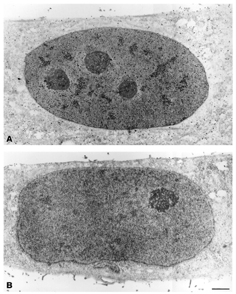
Nucleus and nucleolus, TEM stock photo. Image of cytology ...
Optimized reducing-end labeling of cellulose nanocrystals: Implication ... A strategy to optimize the labeling of the reducing end of native cellulose nanocrystals (CNCs) with gold nanoparticles (AuNPs) was developed and used to investigate the arrangement of the elementary crystallites constituting these biosourced particles. ... Implication for the structure of microfibril bundles in plant cell walls Carbohydr Polym ...
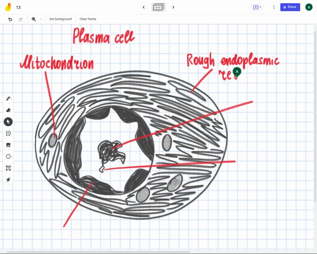
label the transmission electron ricrograph based on the hints provided mitochondnon helerochromalin plasma cell nucleus rough endoplasmic telculn nucleolus 28928
An unlabeled antibody macromolecule technique using hemocyanin for the ... Hemocyanin (Hcy) from whelk, Busycon canniculatum, has been developed as an immunospecific marker for virion and cell surface labeling in the electron microscope. Its size (30 x 50 nm) and distinct cylindrical shape permit easy visualization in the SEM and the transmission electron microscope (TEM). The Hcy method involves the preparation of ...
Transmission Electron Micrograph (TEM) of blood sample ...
Immune response: MedlinePlus Medical Encyclopedia Lymphocytes are a type of white blood cell. There are B and T type lymphocytes. B lymphocytes become cells that produce antibodies. Antibodies attach to a specific antigen and make it easier for the immune cells to destroy the antigen. T lymphocytes attack antigens directly and help control the immune response.

DP Biology: Ultrastructure of cells quiz 1.2
AICE Biology Chapter 1: Plant Cell Electron Micrograph Labeling Start studying AICE Biology Chapter 1: Plant Cell Electron Micrograph Labeling. Learn vocabulary, terms, and more with flashcards, games, and other study tools.
FluoroNanogold: Fluorescence and Electron Micrographs of ...
BacPROTACs mediate targeted protein degradation in ... - Cell Jun 03, 2022 · As seen for pArg and cyclomarin head groups, various molecules that bind to the substrate receptor of the ClpCP protease can be incorporated into a functional degrader. Using cell permeable BacPROTACs, we furthermore demonstrate that recruitment of model proteins to ClpCP leads to selective protein degradation in bacterial cells.
2.3.3 Identify structures from electron micrographs of liver ...
Native Immunogold Labeling of Cell Surface Proteins and Viral ... We describe a method for combining immunogold labeling with cryo-electron microscopy (cryo-EM) and cryo-electron tomography (cryo-ET) of the surface proteins of intact mammalian cells or the surface glycoproteins of assembling and budding viruses in the context of virus-infected mammalian cells cultured on EM grids.
Botrytis tulipae conidium: A. Transmission electron ...
Neuroscience by Dale Purves et al. (eds.) (z-lib.org) - Academia.edu Enter the email address you signed up with and we'll email you a reset link.
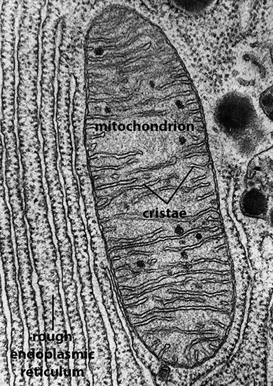
BIOL 230 Lecture Guide - Electron Micrograph of Mitochondria
MicroRNA sequence codes for small extracellular vesicle ... - Nature 22-12-2021 · Exosomes and other small extracellular vesicles (sEVs) provide a unique mode of cell-to-cell communication in which microRNAs (miRNAs) produced and released from one cell are taken up by cells at ...
Transmission Electron Microscopy in Cell Biology: sample ...
Solved Label the transmission electron micrograph of the - Chegg Label the transmission electron micrograph of the cell. 0 Nucleus rences Mitochondrion Heterochromatin Peroxisome Vesicle ULAR bumit Click and drag each label into the correct category to indicate whether it pertains to the cytoplasm or the plasma membrane.

Transmission electron micrographs of armed chlorenchyma cells ...
PDF Identifying Organelles from an Electron Micrograph - Ms JMO's Biology ... The photograph shown below details chloroplast structure as viewed with a transmission electron microscope Courtesy of Dr. Julian Thorpe - EM & FACS Lab, Biological Sciences University Of Sussex A single Granum Chloroplast envelope visible as two membranes Stroma containing numerous small ribosomes Lamellae connecting different grana

Labeling the Cell Flashcards | Quizlet
Immunogold labeling for the diagnosis of leukemia by transmission and ... For the cell type diagnosis of leukemia in adult patients, particularly when the sampling of bone marrow is difficult, the study of peripheral blood leukocytes (PBLs) by immuno-electron microscopy provides significant information, as illustrated here in two cases of hairy cell leukemia and seven cases tentatively identified as megakaryoblastic leukemia (M7).
Multi-color electron microscopy by element-guided ...
Biomimetic material degradation for synergistic enhanced ... - Nature 05-08-2022 · Inefficient tumour treatment approaches often cause fatal tumour metastases. Here, we report a biomimetic multifunctional nanoplatform explicitly engineered with a Co-based metal organic framework ...
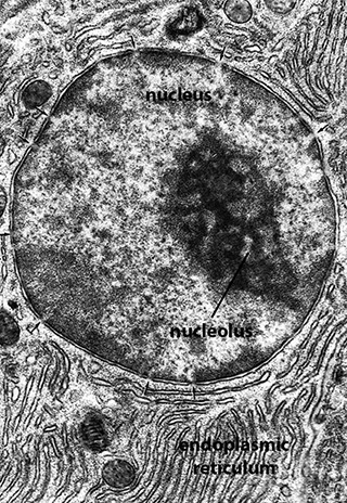
BIOL 230 Lecture Guide - Electron Micrograph of a Nucleus
The Transmission Electron Microscope | CCBER - UC Santa Barbara What is a Transmission Electron Microscope? Transmission electron microscopes (TEM) are microscopes that use a particle beam of electrons to visualize specimens and generate a highly-magnified image. TEMs can magnify objects up to 2 million times. In order to get a better idea of just how small that is, think of how small a cell is.
Electron micrograph plant cell hi-res stock photography and ...
Binding of a Putative and a Known Chaperone Protein Revealed by ... Binding of a Putative and a Known Chaperone Protein Revealed by Immunogold Labeling Transmission Electron Microscopy: A Suggested Use of Chaperones as Probes for the Distribution of Their Target Proteins

transmission electron micrograph of light cells showing ...
Transmission electron microscopy DNA sequencing - Wikipedia Transmission electron microscopy DNA sequencing is a single-molecule sequencing technology that uses transmission electron microscopy techniques. The method was conceived and developed in the 1960s and 70s, but lost favor when the extent of damage to the sample was recognized. In order for DNA to be clearly visualized under an electron microscope, it must be labeled with heavy atoms.
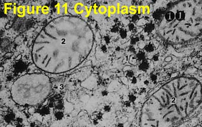
Electron Micrographs
Quantitative investigation of the formation and growth of palladium ... Using liquid-cell transmission electron microscopy, the transformation of irregular palladium nanoclusters to fractal nanocrystals is in situ investigated. The results demonstrate that increasing the difference between the atomic formation/incorporation rates and diffusion rates can promote the formation and growth of dendrites via ...

Transmission electron micrograph (magnification, × 4000). a ...
Animal Cell Electron Microscope Labelled - Q14 Draw a large diagram of ... Here is an electron micrograph of an animal cell with the labels superimposed: Make your work easier by using a label. Make your work easier by using a label. After this, add another oval shape outside the line you just drew, and this will make the cell membrane to your animal cell. You see that many features are in common.

Solved Mitochondrion Nucleus Vesicle Peroxisome | Chegg.com
Ultrasensitive detection of patulin based on a Ag+-driven one … Additionally, from the transmission electron micrograph of AuNFs in Fig. 1E at lower magnification, AuNFs formed a three-dimensional flower-like structure. The UV–vis absorption spectra in Fig. 1 F show an absorbance peak of AuNFs at 622 nm that is not only broader, but has also significantly red-shifted, compared to that of gold nanocrystals at 520 nm.

Transmission electron micrograph (magnification, × 4000). a ...
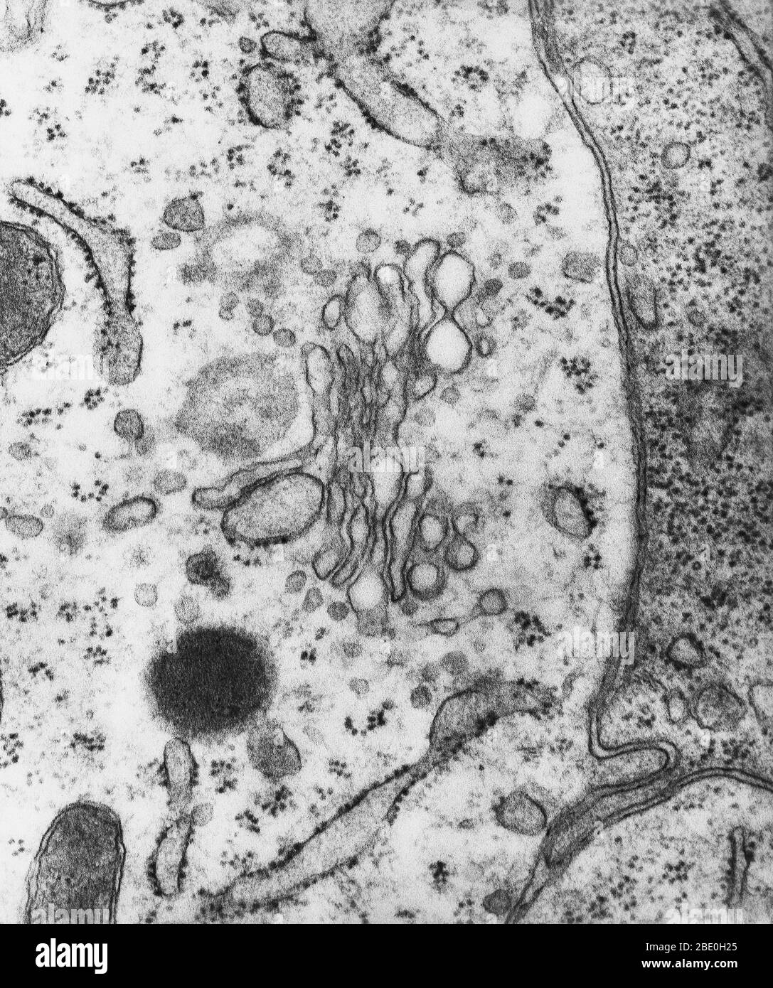
To label: MHC-I, MHC-II, pseudopod and a vesicle in the transmission ... They form invaginations in the outer membrane called as pseudopodia which help in the engulfment of the foreign particles. These foreign antigens are trapped inside the vesicles as evident in electron microscopy. To label: MHC-I, MHC-II, pseudopod and a vesicle in the transmission electron micrograph of a dendritic cell.
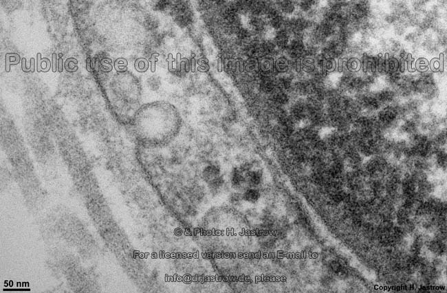
cell and organelles Dr.Jastrow's electron microscopic atlas
anatomy 10.png - Label the transmission electron micrograph... label the parts of the skin. take/launch.jsp? course, assessment_id=_68811_1&course_id= Status: 3 14 2 17 1 13 - 6 12 18 O a Q&A Discuss assumptions of the Transpersonal Caring relationship.

Transmission electron micrograph of an animal cell - Stock ...
Assignment 6, page 2 - North Carolina State University Study this transmission electron micrograph of a spinach leaf cell, locate a chloroplast and capture the image for labeling. The micrograph is displayed as if using a "virtual electron microscope", so you will need to magnify the image and move to a region that contains the clearest view of chloroplast internal structures.

1.2 Skill: Interpretation of electron micrographs - YouTube
Fluorescence microscope - Wikipedia A fluorescence microscope is an optical microscope that uses fluorescence instead of, or in addition to, scattering, reflection, and attenuation or absorption, to study the properties of organic or inorganic substances. "Fluorescence microscope" refers to any microscope that uses fluorescence to generate an image, whether it is a simple set up like an epifluorescence …

Labeling the Cell Flashcards | Quizlet
Electron microscopes - Cell structure - Edexcel - BBC Bitesize the scanning electron microscope (SEM) has a large depth of field. so can be used to examine the surface structure of specimens A human lymphocyte white blood cell as seen with a transmission ...
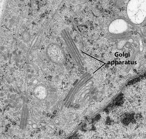
BIOL 230 Lecture Guide - Electron Micrograph of a Golgi Body
Labeling the Cell Flashcards | Quizlet outside the cell wall Label the transmission electron micrograph of the cell. Label the transmission electron micrograph of the mitochondrion. Label the transmission electron micrograph of the nucleus. membrane bound organelles golgi apparatus, mitochondrion, lysosome, peroxisome, rough endoplasmic reticulum nonmembrane bound organelles

2.3.3 Identify structures from electron micrographs of liver ...
Micro Quiz 2 (Chapters 3, 4) Flashcards | Quizlet transmission electron microscope confirming the 9 + 2 microtubule arrangement in a eukaryotic flagellum viewing ribosomes (20 nm) within a bacterial cell viewing a cross section of poliovirus (30 nm)viewing the layers of the gram-negative cell wall in cross section

Solved Label the transmission electron micrograph based on ...
Electron Micrographs of Cell Organelles | Zoology - Biology Discussion This is an electron micrograph of nucleus. (Fig. 17 & 18): (1) Nucleus was discovered by Brown (1831). (2) It is a characteristic entity of almost all eukaryotic cells except mammalian RBCs. (3) The nucleus is generally one but may also be two, four or many.

Transmission electron microscope (TEM) micrograph showing the ...
Immune response: MedlinePlus Medical Encyclopedia Lymphocytes are a type of white blood cell. There are B and T type lymphocytes. B lymphocytes become cells that produce antibodies. Antibodies attach to a specific antigen and make it easier for the immune cells to destroy the antigen. T lymphocytes attack antigens directly and help control the immune response.
Biology, The Cell, Cell Structure, The Endomembrane System ...
A Guide for Using Transmission Electron Microscopy for ...

Transmission electron microscopy images of immunogold-labeled ...
Transmission Electron Microscopy Reveals Distinct Macrophage ...

Cell Micrographs | BioNinja
Transmission Electron Micrograph (TEM) showing mitochondria ...
Transmission electron microscopy cells hi-res stock ...

1.2 Ultrastructure of Cells
Post a Comment for "39 label the transmission electron micrograph of the cell"