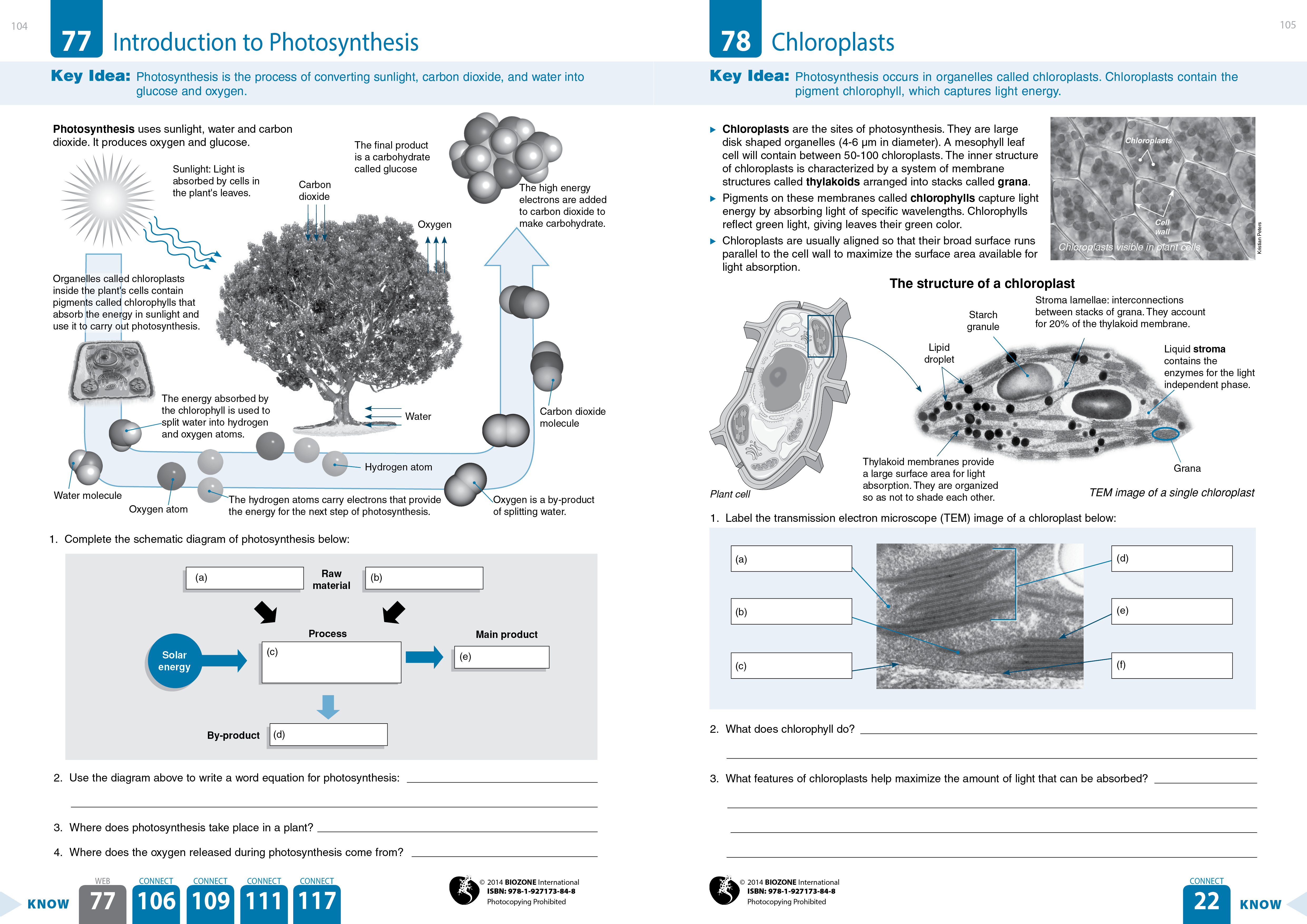
45 label the transmission electron microscope image of a chloroplast below
Labeling the Cell Flashcards | Quizlet Label the transmission electron micrograph of the nucleus. membrane bound organelles golgi apparatus, mitochondrion, lysosome, peroxisome, rough endoplasmic reticulum nonmembrane bound organelles ribosomes, centrosome, proteasomes cytoskeleton includes microfilaments, intermediate filaments, microtubules Identify the highlighted structures Classic | Jailbreak Wiki | Fandom Label The Transmission Electron Microscope Image Of A Chloroplast Below Electron Biogenesis Photosystem Localized Translation; Flying Dragon Easy Sketch Learn How To Draw A Harpy Eagle (bird Of Prey) Step By Step : Drawing; Modern Family Floor Plan Plan Brenna Modern Story Farmhouse Living; Blank Sega Genesis Box Art Minecraft Pc Box Art Cover ...
DP Biology: Ultrastructure of cells quiz 1.2 - ThinkIB The electron microscope image below shows an organelle found in both animal and plant cells. The black line is a scale bar showing 1µm. What is the name of the organelle? A. Mitochondrion B. Chloroplast C. Nucleus D. Rough endoplasmic reticulum Check 3 The electron microscope image below shows an organelle found in both animal and plant cells.

Label the transmission electron microscope image of a chloroplast below
Quia - AP Chapter 6 - Cells (basic) What type of electron microscope produced this image?, transmission electron microscope (TEM), The cell below is a(n) _____ cell., animal, The cell below is a(n) _____ cell., plant, The ____ is the simplest collection of matter that can live. cell: The first microscopes, as well as the microscopes that we use in lab, are called ____. light ... PDF Scanned Document - Bronx High School of Science Study the electron micrographs in your text. Describe the different types of images obtained from: scanning electron microscopy (SEM) transmission electron microscopy (T EM) In cell fractionation, whole cells are broken up in a blender, and this slurry is centrifuged several times. Each time, smaller and smaller cell parts are isolated. Plant Cell Nucleus Electron Micrograph : Cell And Organelles Dr Jastrow ... In electron micrographs, centrioles appear as cylindrical structures which occur in pairs lying at right angles to each other (figs. Chloroplast Wikipedia from upload.wikimedia.orgThis electron micrograph is higher in magnification. You see that many features are in common. Typically, the nucleus is the most prominent organelle in a cell.
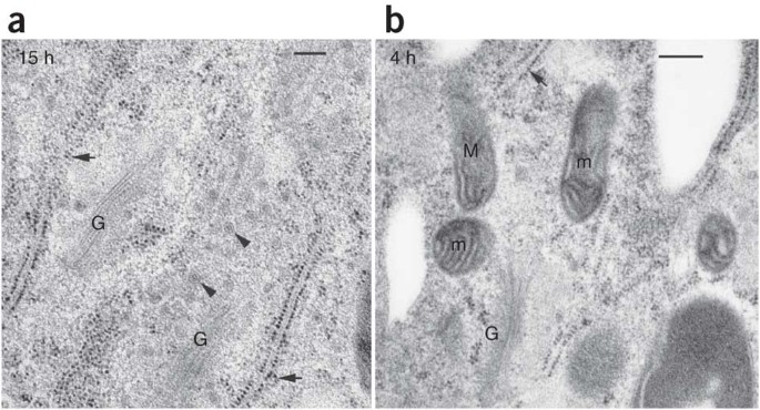
Label the transmission electron microscope image of a chloroplast below. Cells and magnification Flashcards - Quizlet 6) a- Figure 2 shows a photos of part of a mitochondrion from a mouse liver cell taken using a transmission electron microscope at x62800 magnification Produce a scientific drawing of the mitochondria in figure 2 Label the following part of the mitochondrion on your drawing -Matrix-Crista (4 marks) Assignment 6, page 1 - NC State University View this transmission electron micrograph of a plant cell, locate a chloroplast and capture the image for labeling. The micrograph is displayed as if using a "virtual electron microscope", so you will need to magnify the image and move to a region that contains the clearest view of chloroplast internal structures. Amazing 27 Things Under The Microscope With Diagrams The tail is transparent and thus is difficult to detect under a low-power microscope. 23. Spirogyra under the microscope. Spirogyra is a green alga found mostly in freshwater in the form of green clumps. Spirogyra is unicellular, but because it clumps together, it can be seen in the pond even with our naked eyes. Transmission electron microscopic images of chloroplasts and ... Transmission electron microscopic images of chloroplasts and mitochondria in 15-day-old leaves from PRORP1 RNAi mutants and wild-type plants. (A, B) Ultrastructure of chloroplasts and mitochondria...
Worksheet microscopes and cells.pdf - Course Hero • A scanning/transmission electron microscope beams electrons at the surface of a 3D object. • A higher power lens increases/decreases the size and detail of the image of the specimen. • To calculate the total magnification, multiply/divide the power of the objective lens with the ocular lens. PDF MISSION Engage students to continuously learn, Fall 2021 Describe the different types of images obtained from and limitations to: (a) scanning electron microscopy (SEM) (b) transmission electron microscopy (TEM) 3. What is resolving powerand why is it important in biology? 4. In cell fractionation, whole cells are broken up in a blender, and this slurry is centrifuged several times. Chloroplast - Wikipedia Transmission electron micrograph of Chlamydomonas reinhardtii, a green alga that contains a pyrenoid surrounded by starch. Helicosporidium is a genus of nonphotosynthetic parasitic green algae that is thought to contain a vestigial chloroplast. (Get Answer) - Electron micrograph of a plant cell with chloroplasts in ... 5. When a transmission electron microscope is used, cells are usually studied using electron micrographs, photographs taken of the image seen on the fluorescent screen. Observe the electron micrographs in Figure 2.5a and b, respectively, a plant cell and an animal cell.
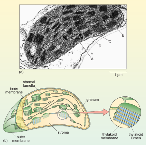
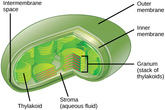
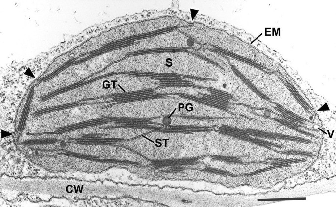
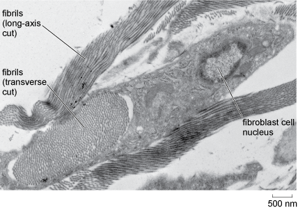
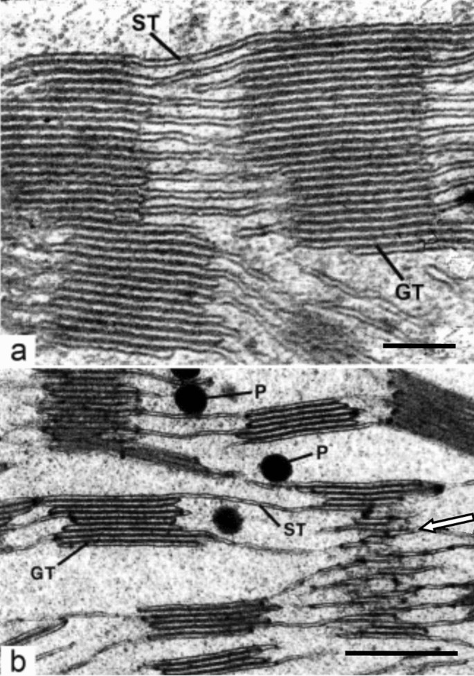
PDF Identifying Organelles from an Electron Micrograph The photograph shown below details chloroplast structure as viewed with a transmission electron microscope Courtesy of Dr. Julian Thorpe - EM & FACS Lab, Biological Sciences University Of Sussex A single Granum Chloroplast envelope visible as two membranes Stroma containing numerous small ribosomes Lamellae connecting different grana Classical transmission electron microscopy (TEM) led to the formulation ... Thin section electron micrograph of a chemically fixed chloroplast in a young tobacco leaf. The chloroplast lies flat against the plasma membrane and the cell wall (CW) and presents a more or less elliptical outline. The stacked grana thylakoids (GT) are interconnected by non-stacked stroma thylakoids (ST). Electron microscopes - Cell structure - Edexcel - BBC Bitesize the transmission electron microscope (TEM) is used to examine thin slices or sections of cells or tissues the scanning electron microscope (SEM) has a large depth of field so can be used to examine... Looking at the Structure of Cells in the Microscope With a normal bright-field microscope, the image is obtained by the simple transmission of light through a cell in culture. Images of the same cell obtained by four kinds of light microscopy are shown in Figure 9-8. Figure 9-8 Four types of light microscopy. Four images are shown of the same fibroblast cell in culture.
Transcriptomic and proteomic analyses reveal new insight into ...
DP Biology (Cells: 1.1, 1.2, 1.5, 6.3) Quiz - Quizizz The image shows a phagocytic white blood cell as seen with a transmission electron microscope. Which features can be found both within this cell and in a photosynthetic bacterium? answer choices chloroplasts multiple nuclei 70s ribosomes lysosomes Question 2 30 seconds Q. The image shows an electron micrograph of a fungus, Candida albicans.

Chloroplast - Wikipedia
Electron Microscope- Definition, Principle, Types, Uses, Labeled Diagram There are two types of electron microscopes, with different operating styles: 1. Transmission Electron Microscope (TEM) The transmission electron microscope is used to view thin specimens through which electrons can pass generating a projection image. The TEM is analogous in many ways to the conventional (compound) light microscope.

Cell Micrographs | BioNinja
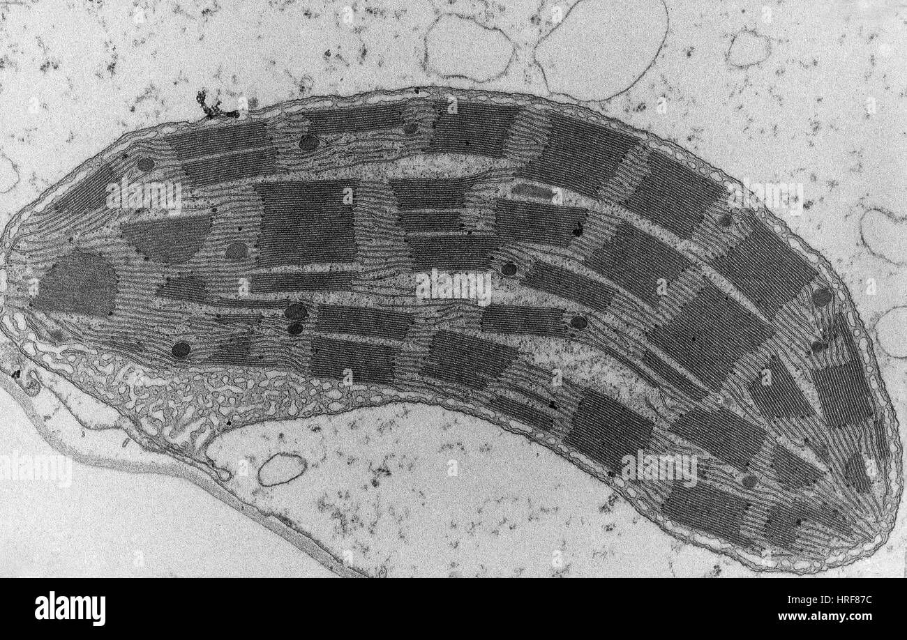
Electron Micrographs of Cell Organelles - Biology Discussion This is an electron-micrograph of plastid or chloroplast, which is an integral component of all green plant leaves and is characterized by following features (Fig. 15 & 16): (1) They may be spheroidal, ovoid, stellate or collar shaped and differ in size and number in different cells.
Preparation of plant cells for transmission electron ...
Cell and Molecular Biology part 1 Flashcards - Cram.com Draw and label a simple diagram of a phospholipid molecule (2) Why are the phospholipid molecules arranged in a bilayer? ... the chloroplast grana. B) the mitochondrial matrix . C) the chlproplast stroma. ... Transmission electron microscope images of bacteria show that they also have a nucleus surrounded by s nuclear envelope, but lack ...
f i1w,-
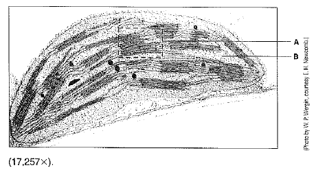
Quia - AP Chapter 6 - Cells (detailed) What type of electron microscope produced this image?, transmission electron microscope (TEM), The cell below is a(n) _____ cell., animal, The cell below is a(n) _____ cell., ... The "A" is pointing to ____ in the picture below., chloroplast DNA (both chloroplasts and mitochondria have DNA of their own. It's thought to be strong evidence of the ...
A tour of the cell: View as single page
(374135792) Chapter 6 Tour of the Cell - DocShare.tips The development of electron microscopes has further opened our window on the cell and its organelles. What is considered a major disadvantage of the electron microscopes? Cell dies in the process 3. Study the electron micrographs in your text. Describe the different types of images obtained from: scanning electron microscopy (SEM)
What is a diagram of a plant and animal cell under an ...
What are the labels of the transmission electronic microscope image of ... Transfer RNA (tRNA) precursors undergo endoribonucleolytic processing of their 5' and 3' ends. 5' cleavage of the precursor transcript is performed by ribonuclease P (RNase P). While in most organisms RNase P is a ribonucleoprotein that harbors a catalytically active RNA component, human mitochondria and the chloroplasts (plastids) and mitochondria
A unifying structural and functional model of the coronavirus ...
Electron Microscopy Images - Dartmouth Transmission electron microscope image of a thin section cut through the bronchiolar epithelium of the lung (mouse), which consists of ciliated cells and non-ciliated cells. Image shows the ciliary microtubules in transverse and oblique section. In the cell apex are the basal bodies that are the anchoring sites for the cilia.
3.3 Eukaryotic Cells – Concepts of Biology – 1st Canadian Edition
The Transmission Electron Microscope | CCBER Transmission electron microscopes (TEM) are microscopes that use a particle beam of electrons to visualize specimens and generate a highly-magnified image. TEMs can magnify objects up to 2 million times. In order to get a better idea of just how small that is, think of how small a cell is.

What are the labels of the transmission electronic microscope ...
Part 3. Structure of The Plant Leaf and Chloroplasts Study this transmission electron micrograph of a spinach leaf cell, locate a chloroplast and capture the image for labeling. The micrograph is displayed as if using a "virtual electron microscope", so you will need to magnify the image and move to a region that contains the clearest view of chloroplast internal structures.
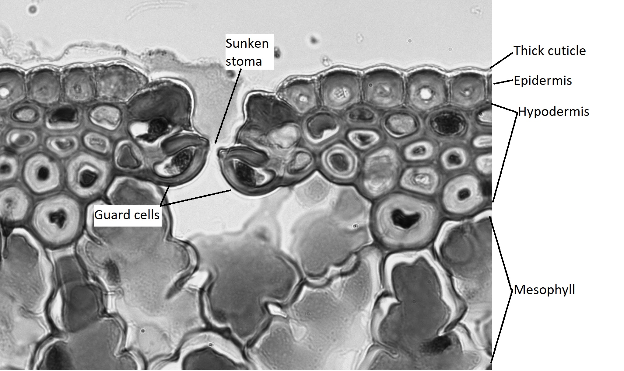
12.2: Internal Leaf Structure - Biology LibreTexts
cells ESQ (1).doc - Course Hero Feature Mitochondrion Chloroplast Double outer membrane Starch grains Diffusion of oxygen into the organelle (3) (c) ... Contrast how an optical microscope and a transmission electron microscope work and contrast the limitations of their use when ... The image below shows the cell-surface membrane of a red blood cell seen with a transmission ...
30 Label The Transmission Electron Microscope Image Of A ...
Plant Cell Nucleus Electron Micrograph : Cell And Organelles Dr Jastrow ... In electron micrographs, centrioles appear as cylindrical structures which occur in pairs lying at right angles to each other (figs. Chloroplast Wikipedia from upload.wikimedia.orgThis electron micrograph is higher in magnification. You see that many features are in common. Typically, the nucleus is the most prominent organelle in a cell.

Materials | Free Full-Text | Effect of Third-Stage Heat ...
PDF Scanned Document - Bronx High School of Science Study the electron micrographs in your text. Describe the different types of images obtained from: scanning electron microscopy (SEM) transmission electron microscopy (T EM) In cell fractionation, whole cells are broken up in a blender, and this slurry is centrifuged several times. Each time, smaller and smaller cell parts are isolated.
f i1w,-
Quia - AP Chapter 6 - Cells (basic) What type of electron microscope produced this image?, transmission electron microscope (TEM), The cell below is a(n) _____ cell., animal, The cell below is a(n) _____ cell., plant, The ____ is the simplest collection of matter that can live. cell: The first microscopes, as well as the microscopes that we use in lab, are called ____. light ...

Overexpression of the chloroplastic 2-oxoglutarate/malate ...
Chloroplast micrograph hi-res stock photography and images ...
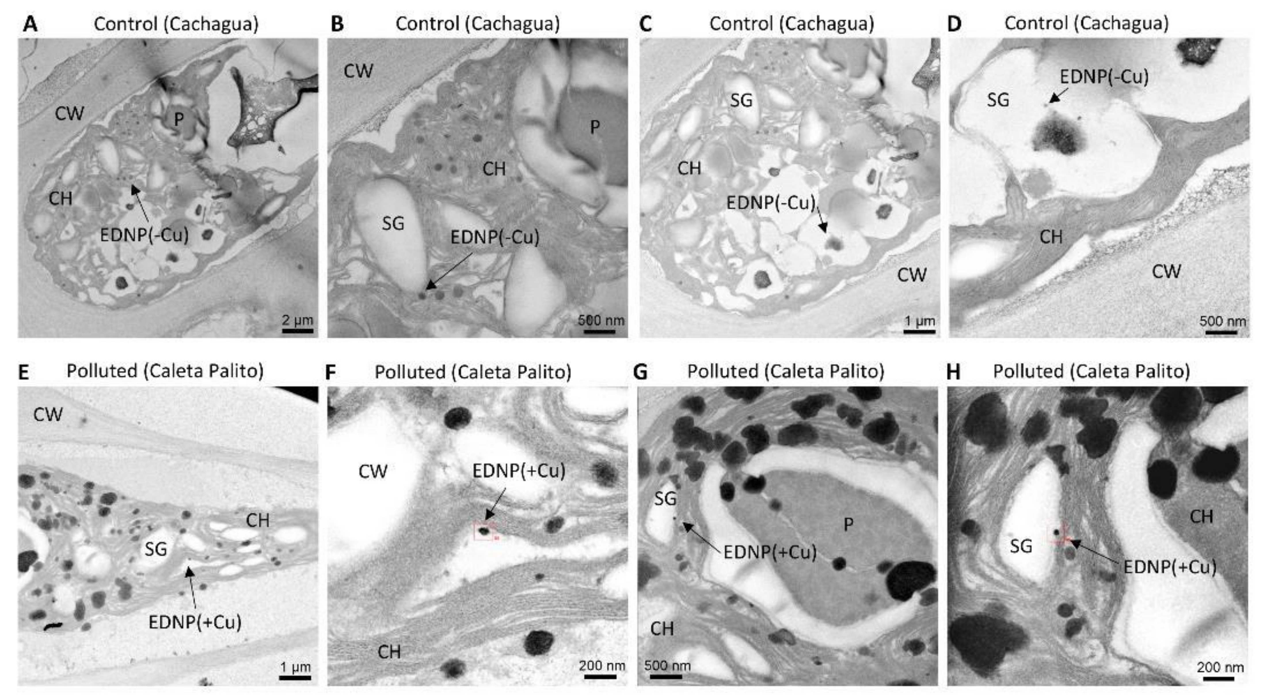
IJMS | Free Full-Text | Ulva compressa from Copper-Polluted ...

Transmission electron microscopy picture of the chloroplast ...

Plastoglobuli, Thylakoids, Chloroplast Structure and ...

Bio 150 - Microscopy and Cell Structure (Lab 5) Flashcards ...

Vivian_Slack_-_ ...

Chloroplast
What is a diagram of a plant and animal cell under an ...
A brief history of how microscopic studies led to the ...

Penetration of sub-micron particles into dentinal tubules ...
WHIRLY1 functions in the nucleus to regulate barley leaf ...

Transmission electron micrographs of immunogold labelling ...
A tour of the cell: View as single page

Ion Pathways in Biomineralization: Perspectives on Uptake ...
What cell organelles can be seen under the electron ...

Assignment 6, page 2

Chloroplast Sec14-like 1 (CPSFL1) is essential for normal ...
IJMS | Free Full-Text | Subcellular Sequestration and Impact ...

Photosynthesis Problem Set 1

PDF) A primer on resolving the nanoscale structure of the ...
Solved Examine this electron micrograph of a chlorplast ...

Peptide-mediated Targeting of Nanoparticles with Chemical ...
A brief history of how microscopic studies led to the ...

Transmission Electron Microscopy of Biological Samples ...
Enhanced Survival of the Cyanobacterium
Post a Comment for "45 label the transmission electron microscope image of a chloroplast below"